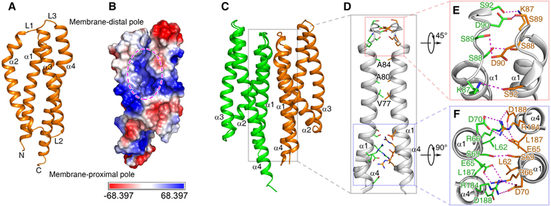
Fig. 1. The structure of ligand-free MCP2201LBD.

Cartoon (A) and electrostatic potential surface (B) representation of a monomer, the dimeric structure (C) and its interface (D) are shown. The ligand binding pocket in (B) is highlighted with ellipse. Two major parts of dimer interface, the membrane-distal and membrane-proximal ones, are enlarged in panels (E) and (F).
