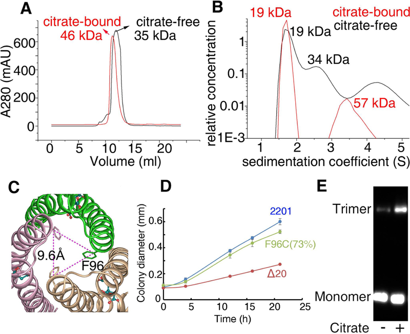
Fig. 4. Size-exclusion chromatography, analytical ultracentrifugation and TMEA crosslinking assays support a trimeric structure of citrate-bound MCP2201LBD.

(A) Size-exclusion chromatography assay of MCP2201 LBD. (B) Analytical ultracentrifugation assay of MCP2201 LBD. For (A) and (B), the plots of proteins in the absence and presence of citrate are colored as black and red, respectively. (C) Top view of citrate-bound trimer-of-monomers structure of MCP2201 LBD showing F96 location. (D) The plots of swimming ring diameter vs time for strain harboring MCP2201 F96C mutant. (E) TMEA crosslinking of integral MCP2201 variant F96C. The bands of monomer and trimer are indicated.
