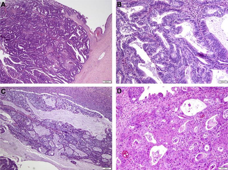
Figure 1. Histologic features of endometrioid adenocarcinomas of the ovary.
(A) Well-differentiated endometrioid adenocarcinoma of the ovary arising in a background of endometriosis. (B) Higher power micrograph of a well-differentiated endometrioid ovarian carcinoma. (C) Well-differentiated endometrioid adenocarcinoma of the ovary with mucinous differentiation. (D) Well-differentiated endometrioid ovarian carcinoma with squamous differentiation. Scale bars, 500 μm.

