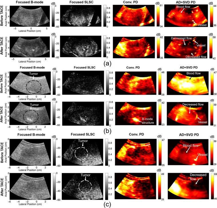
Figure 2.
Focused B-mode, focused SLSC, and power Doppler images before and after TACE for subjects 10 (a), 9 (b), and 5 (c). Power Doppler (PD) images were made with conventional (Conv.) methods and with adaptive demodulation (AD) and SVD filtering. 27 ms (16 samples) and 0.5 s (300 samples) ensemble sizes were used for the conventional and advanced methods, respectively. SLSC images are scaled from 0 to the maximum coherence in the image. Potential vessels, tumor flow, and decreased flow are indicated in the AD + SVD images that are not apparent in the conventional PD images.

