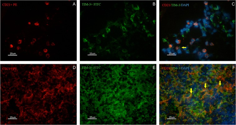
Figure 6.
Identification of CD21+ and TIM-3+ cells by immunofluorescence in the spleen of dogs naturally infected by L. infantum presenting organized (A–C) or disorganized (D–F) splenic white pulp. In (A,D) the presence of CD21+ cells/B lymphocytes (red PE) is shown. In (B,E) the presence of TIM-3+ cells (green FITC) is shown. In (C,F) overlapping images (red PE/green FITC/blue DAPI) are shown. Yellow arrows indicate CD21+ cells also expressing TIM-3+. Scale bar: 25 μm.

