Figure 1.
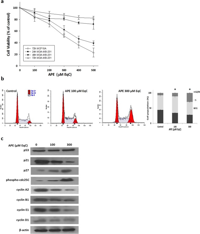
APE inhibits MDA-MB-231 cell growth and induces G2/M phase arrest. (a) Effect of APE on MDA-MB-231 and MCF10A cell viability. MDA-MB-231 and MCF10A cells were cultured for 24, 48, and 72 h in medium supplemented or not (control) with APE 100, 200, 300, 400, and 500 μM EqC. Cell viability was then assessed by MTT assay and expressed as a percentage of untreated cells. Values represent the mean ± SD of three independent experiments. (b) MDA-MB-231 cells were treated with APE 100 and 300 μM EqC for 24 h. The distribution of cell cycle was assessed by flow cytometry. PI fluorescence was collected as FL3-A (linear scale) by the ModFIT software (Becton Dickinson). For each sample at least 2 × 10⁴ events were analyzed in at least three different experiments giving a SD less than 5% (*P < 0.05 versus control). (c) The levels of cell cycle-regulatory proteins in MDA-MB-231 cells treated with APE 100 and 300 μM EqC for 24 h were measured by western blotting. β-actin was used as a standard for the equal loading of protein in the lanes. The full-length blots are included in the supplementary information (Fig. S1).

