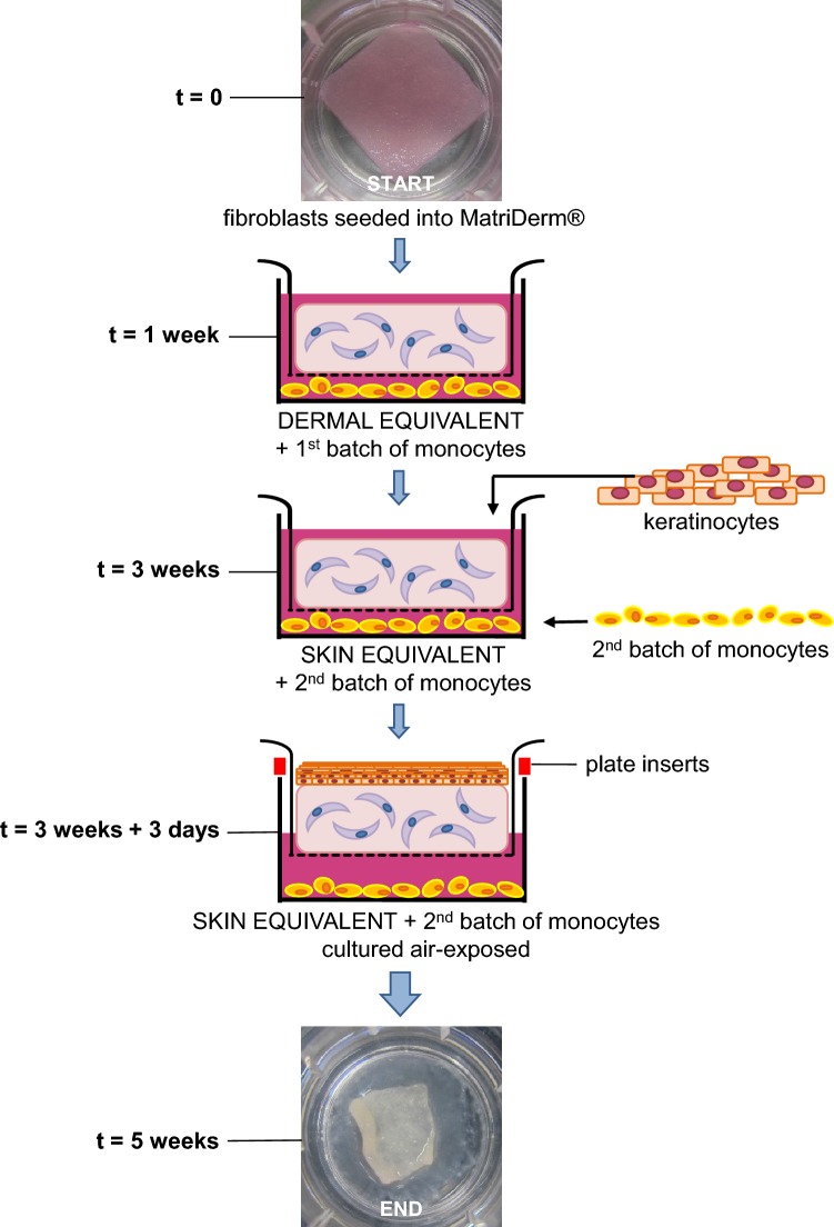
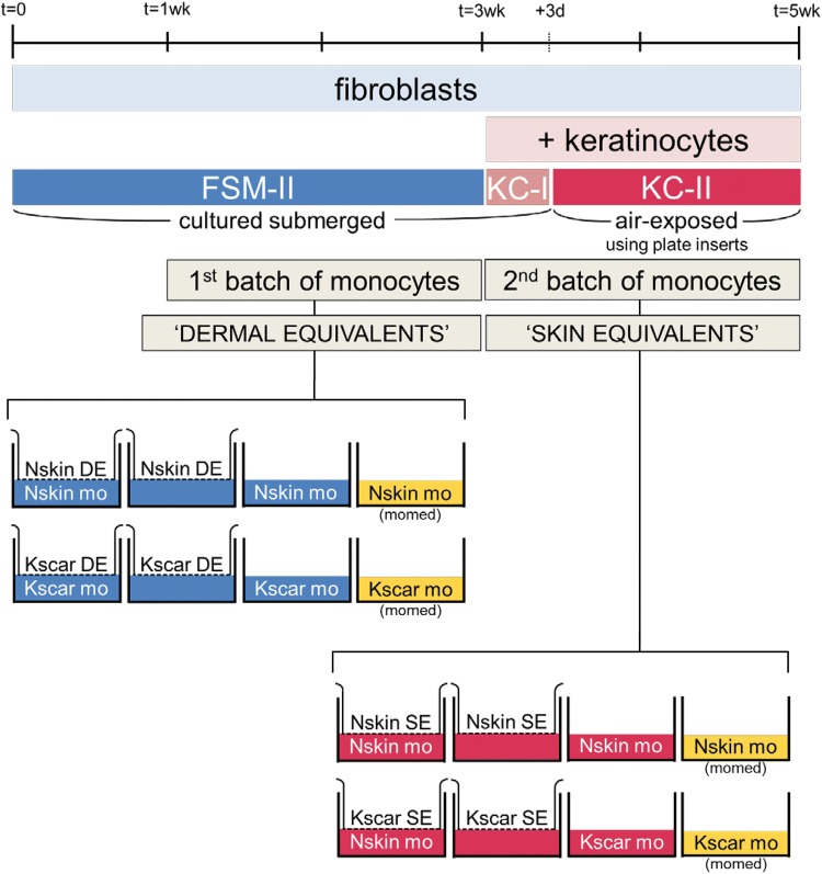
Fig. 1.
Construction of skin models. a Skin models are constructed by first seeding fibroblasts into MatriDerm® (first picture: macroscopic view). After 3 weeks, keratinocytes are added and the skin models are then cultured air-exposed for an additional 2 weeks. The first batch of monocytes was added during week 1–3 (only fibroblasts present, DE stage) and the second fresh batch of monocytes during week 3–5 (fibroblasts and keratinocytes present, SE stage). The last macroscopic image shows the same skin model as shown above, at 5 weeks after initiating culture. The diameter of the transwell insert is 24 mm. b Overview of the culturing process during the entire 5-week timeline as explained in ‘Materials and methods’. The first batch was co-cultured with DE and second monocyte batch was co-cultured with SE. The experimental conditions for both stages are depicted in the final row. From left to right, Nskin/Kscar skin models were co-cultured with their respective monocytes for 2 weeks (SE/DE + mo) and compared to the three control conditions: skin models only (SE/DE), monocytes only cultured in either the same medium as the skin models (mo) or in their own medium (mo, mo-medium). wk week, d days, DE dermal equivalents, SE skin equivalents, mo monocytes, Nskin normal skin, Kscar keloid scar, mo-medium monocyte medium, FSM-II, KC-I and KC-II: various types of media used, see supplementary Table 1


