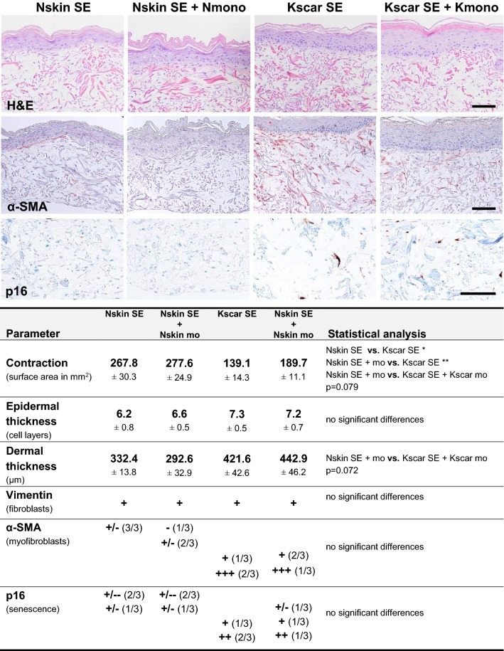
Fig. 2.
Effect of monocyte co-culture on the keloid scar phenotype. Images show immunohistochemical staining of normal skin equivalents cultured alone (Nskin SE) or with monocytes (Nskin SE + mo), and keloid skin equivalents cultured alone (Kscar SE) or with monocytes (Kscar SE + mo). The table summarizes the results for n = 3 normal skin (Nskin) and n = 3 keloid scars (Kscar), with or without monocytes (mo): contraction was measured as a reduction in the end surface area after a 5-week culture; epidermal thickness was measured as the number of viable epidermal cell layers in the SE; dermal thickness was measured in μm; vimentin staining indicated the presence fibroblasts within the MatriDerm®; α-SMA stained myofibroblasts; p16 was used as a senescence marker. Scoring legend for immunohistochemical staining: ± minimal expression, +: normal expression, ++ increased expression, +++ strongly increased expression, − absent. Results in the table were shown as mean ± SEM; vs.: versus (compared to). An ordinary one-way ANOVA with post hoc Tukey’s multiple comparisons test was performed with *p < 0.05, **p < 0.01. Bar = 100 µm

