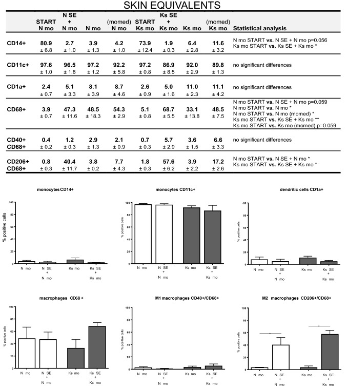
Fig. 4.
Immunophenotyping of monocytes (co-) cultured with full skin equivalents: monocyte, dendritic cell and macrophage marker expression. Monocytes, both mono- and co-cultured with skin equivalents, were analysed for monocyte (CD14+, CD11c+), dendritic cell (CD1a+), macrophage (CD68+), M1 macrophage (CD40+) and M2 macrophage (CD206+) marker expression via FACS analysis. The table summarizes the results of each experimental group by listing the mean % positive staining with SEM with n = 3 for all four normal skin conditions and n = 3 for all four keloid scar experimental groups; vs.: versus (compared to). Statistically significant results of an ordinary one-way ANOVA or Kruskal–Wallist test with post hoc testing on selected groups are listed in the table. Graphs of the results summarized in the table can be found in the right-side columns of supplemental Fig. 4, and the associated figure legends also list the statistical test used for each graph. The lower half of the figure shows the most relevant comparison in graphs: monocytes mono-cultured vs. monocytes co-cultured with Nskin/Kscar models. An ordinary one-way ANOVA with Tukey’s multiple comparisons was performed for CD14, CD11c, CD68, CD40/CD68 and CD206/CD68. The Kruskal–Wallist test with Dunn’s multiple comparison test was performed for CD1a. *p < 0.05, **p < 0.01, ****p < 0.0001. Nskin normal skin, Kscar keloid scar, mo monocytes, momed monocytes cultured in monocyte medium (for contents, see supplemental Table 1). SE skin equivalent comprising keratinocytes forming an epidermal layer on top of fibroblast-populated MatriDerm®; second batch of monocytes co-cultured with full skin equivalent from t = week 3 to week 5

