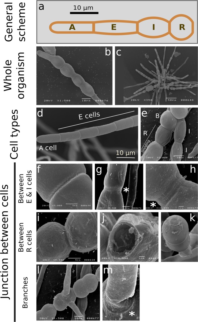
Figure 1.
Filament organisation and cell morphologies observed by scanning electronic microscopy. (a) Overview of Ectocarpus sporophyte filament (prostrate) growing from spore germination. Five cell types are defined according to their position and shape. A type: Apical cell; E type: Elongated, cylindrical cell; I type: Intermediate cell; R type: Round, spherical cells positioned at the central region of the filaments; B type: Branched cells. Cell types are defined according to their position (for A cells) and their ratio of their length (L) to their width (w) (E, I and R cells). E cell: L/w > 2; I cell: L/w in [1.2; 2[; R cell: L/w < 1.2. The number of E, I, R and B increases with the filament maturation stage. Cells of the same cell types are contiguous. (b,c) Whole organism observed by scanning electronic microscopy (SEM); One week post germination (b), or 2–3 weeks post germination (c).(d) A and E cells at the filament extremity. (e) I and R cell types in the central region of the filament. B indicates branching cells. (f–h) Junctions between E cells (f) and I cells (g,h), showing either single- (f) or double- ring(s) framing the wall (asterisks in g, h). (i–k) Junctions between R cells. (l–m) Higher magnification on branches, showing a ring at the junction site (asterisk).

