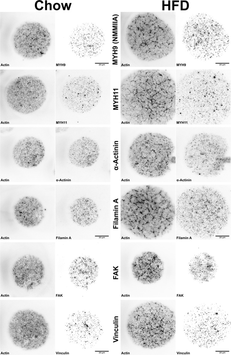
Figure 2.
Representative TIRF images of isolated adipocytes from chow or HFD-fed mice (chow, HFD) that were co-stained with phalloidin to detect filamentous actin and different markers of stress fibers, actin organization and focal adhesions (non-muscle myosin (NMM) IIA isoforms MYH9 and MYH11, α-actinin, Filamin A, FAK, Vinculin). Each marker (black) shown in right panel for each condition, and phalloidin (black) shown in left panel for each condition. Scale bar = 20 µm.

