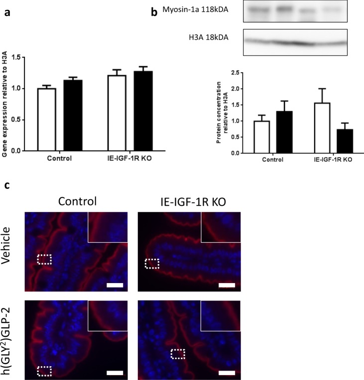
Figure 8.
Myosin-1a remains unchanged with both GLP-2 treatment and IE-IGF-1R KO. Control and IE-IGF-1R KO mice were treated with either vehicle (open bars) or h(GLY2)GLP-2 (closed bars) for 11 d, followed by jejunal mucosal scrapes for either (a) q-RT-PCR or (b) western blot analysis; representative blots are shown, with full length blots in Supplemental Fig. 5 (n = 8–14 for PCR; n = 4–9 for western blot). (c) Jejunal sections were paraffin embedded for immunofluorescent staining; insets show magnified views of boxed areas. Red is myosin-1a, blue is DAPI. Scale bar is 20 µm (n = 4).

