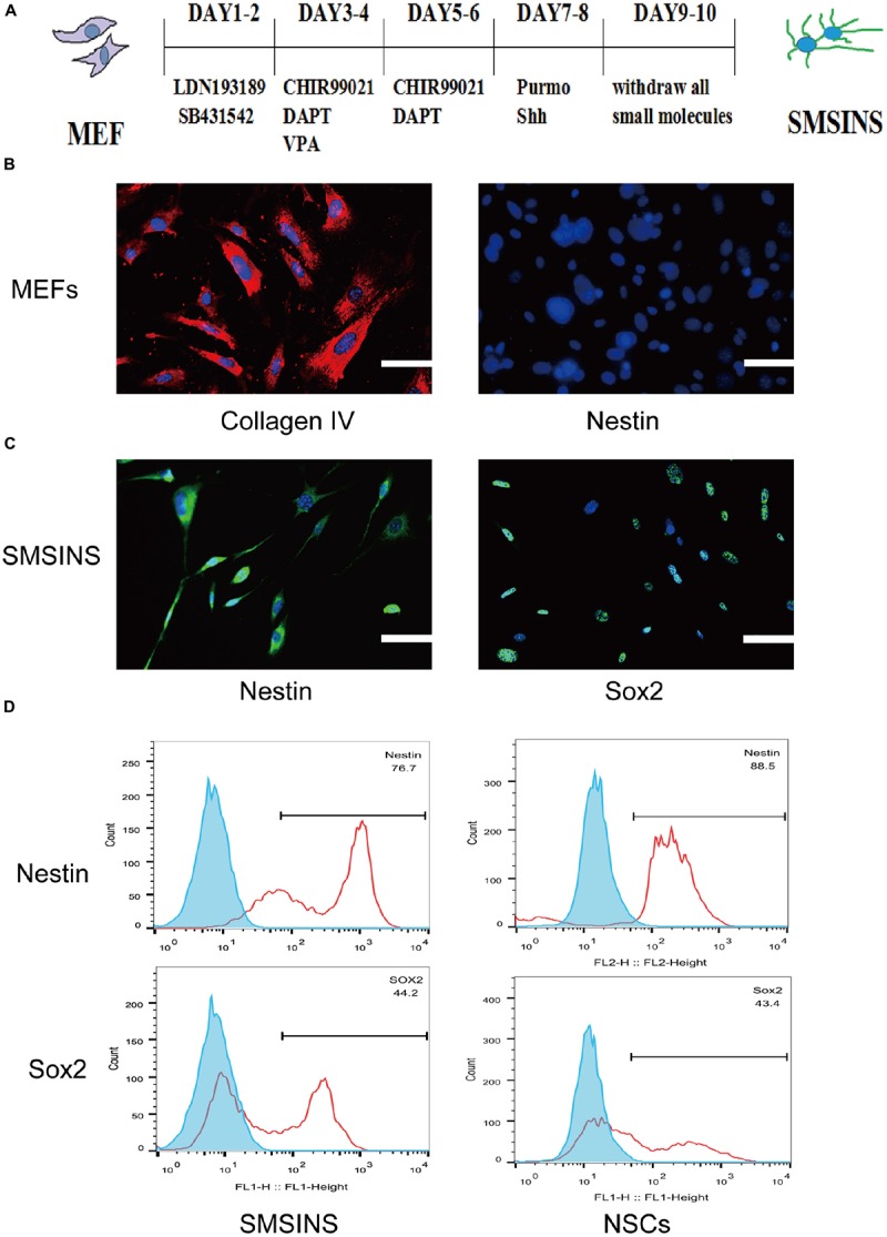
FIGURE 1.

Induction of SMSINS from MEFs. (A) Schematic diagram of the SMSINS induced process. (B) MEFs express fibroblast markers without NSC markers. MEFs were stained with specific antibodies against Collagen IV (Red) and Nestin (Green). (C) Immunostaining of Nestin (Green) and Sox2 (Green) in SMSINS at day 10 using a specific antibody. Nuclei were counterstained with DAPI (blue). (D) Flow cytometric analysis to quantify cells expressing Sox2 and Nestin after SMSINS induction and native NSC cells isolated from embryonic mouse brain. Scale bars: 100 μm (B,C).
