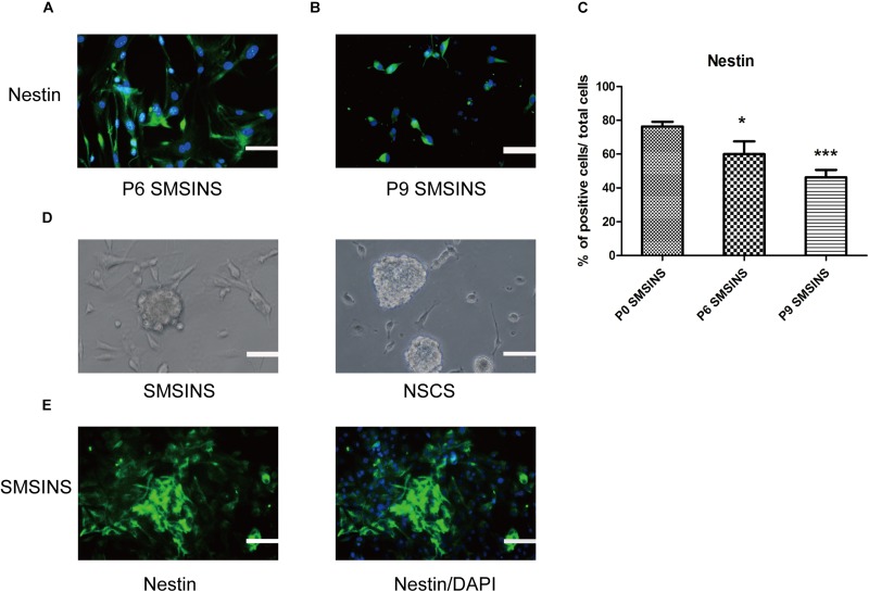
FIGURE 2.
SMSINS expressed NSC markers similar to NSCs. (A) Neuroepithelial-like colonies of SMSINS at passage 6 (P6) stained with a specific antibody against the NSC marker Nestin (Green). (B) Immunostaining of Nestin (Green) in SMSINS at passage 9 (P9). Nuclei were counterstained with DAPI (blue). (C) Quantitative analyses of the protein expression level of Nestin in SMSINS at passage 0, 6, and 9. Data represent the mean ± SEM. F = 12.39 ∗p < 0.05; ∗∗∗p < 0.001 compared with P0 SMSINS; One-way ANOVA followed with Dunnett’s T3 test. (D) SMSINS and native NSCs formed neural spheres when cultured in suspension. (E) Neural spheres of SMSINS stained with specific antibodies against Nestin (Green). Nuclei were counterstained with DAPI (blue). Scale bars: 100 μm.

