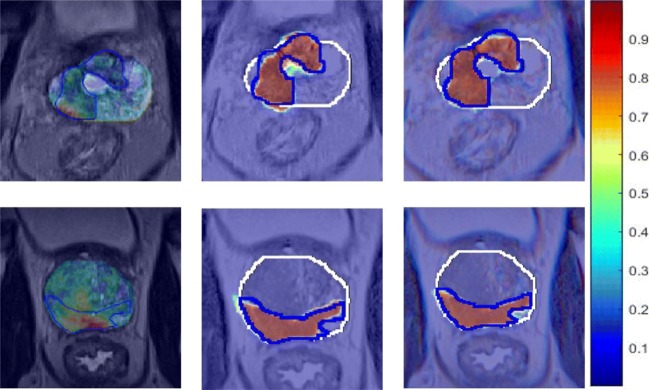
Fig. 4.
Results of prostate cancer detection produced by Lemaitre et al. [21], M1 and M2 from left to right, respectively. White contour shows the prostate boundary segmented by a radiologist, while blue contour is the ground truth of malignant lesions. Note that each row shows the same slice and each column shows the performance of the same CAD system

