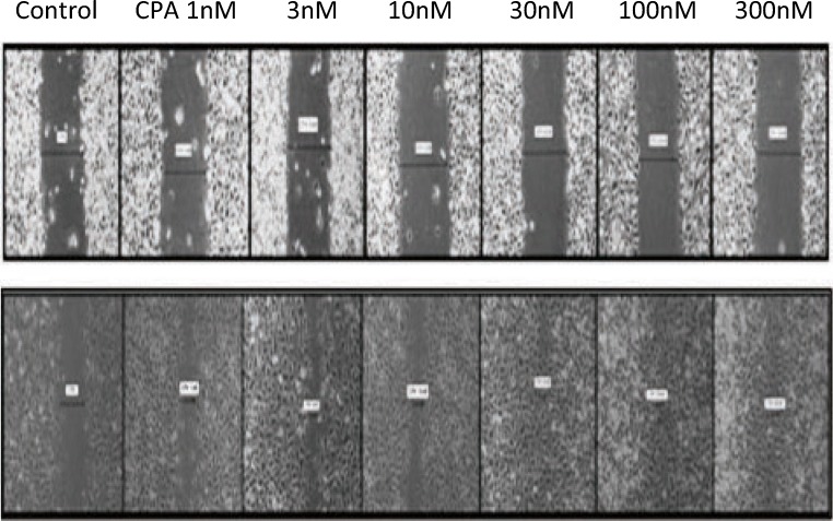
Fig. 2.
Wound healing scratch assay using different concentrations of CPA on EA.hy926 endothelial cells. Upper panel depicts scratch widths at 0 times and lower panel shows scratch widths after 6 h. CPA concentrations from the left to right are control (0 nM), 1 nM, 3 nM, 10 nM, 30 nM, 100 nM and 300 nM CPA. Magnification, × 40

