Figure 4.
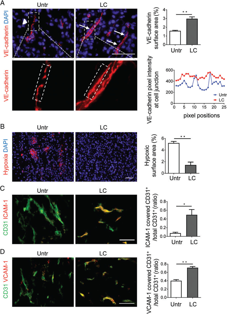
LIGHT–CGKRK treatment improves endothelial barrier integrity and vessel function. (A) NFpp10-GBM mice were left untreated (Untr) or treated for 2 weeks with LIGHT–CGKRK (LC). VE-cadherin expression in tumours was quantitatively (upper) and qualitatively (lower) assessed by IHC. Arrowheads point to gaps in disjointed adherens junctions, and arrows point to continuous VE-cadherin+ adherens junctions; n=3, **p =0.0032. Dashed outlines (grey, upper) indicate the areas enlarged below, and dashed white lines (below) indicate junctional areas quantified as VE-cadherin fluorescence (in pixels, same exposure for all groups) at various positions (horizontal axis) along the cell adherens junctions. (B) Assessment and quantification of intratumoural hypoxia by immunohistological detection of pimonidazole-positive areas (red) in treatment groups; n=3, **p =0.0028. (C and D) Expression of ICAM-1 (C) or VCAM-1 (D) on GBM tumour vessels with and without LC treatment, analysed by IHC and quantified; n=3, *p =0.027, **p =0.0014. Student’s t-test. Scale bars: 50 μm.

