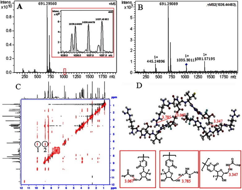
Figure 4.
(A) FT-MS spectrum gives RGDV-gemcitabine an ion peak of monomer and an ion peak of trimer; (B) qCID spectrum of the trimer gives an ion peak of monomer and an ion peak of dimer; (C) NOESY 2D 1H NMR spectrum gives two cross-peaks that defines the approach manner of RGDV-gemcitabine forming trimer; (D) to fit the NOESY 2D 1H NMR spectrum, the trimer of RGDV-gemcitabine should possess butterfly-like conformation.
Abbreviations: RGDV-gemcitabine, 4-(Arg-Gly-Asp-Val-amino)-1-[3,3-difluoro-4-hydroxy-5-(hydroxylmethyl)oxo-lan-2-yl]pyrimidin-2-one.

