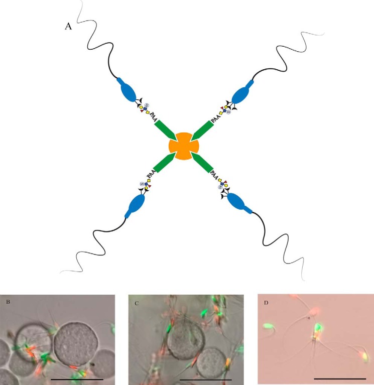
Figure 14.
Sperm bind to suLeA-beads. A schematic shows how biotinylated suLeA or laminin were attached to streptavidin-agarose beads (A). Sperm that bound to suLeA-beads (B) or laminin-beads (C), or were free-swimming (D) were incubated for 8 h and labeled with SYBR14 (green, live) and PI (red, dead). Scale bar, 50 μm.

