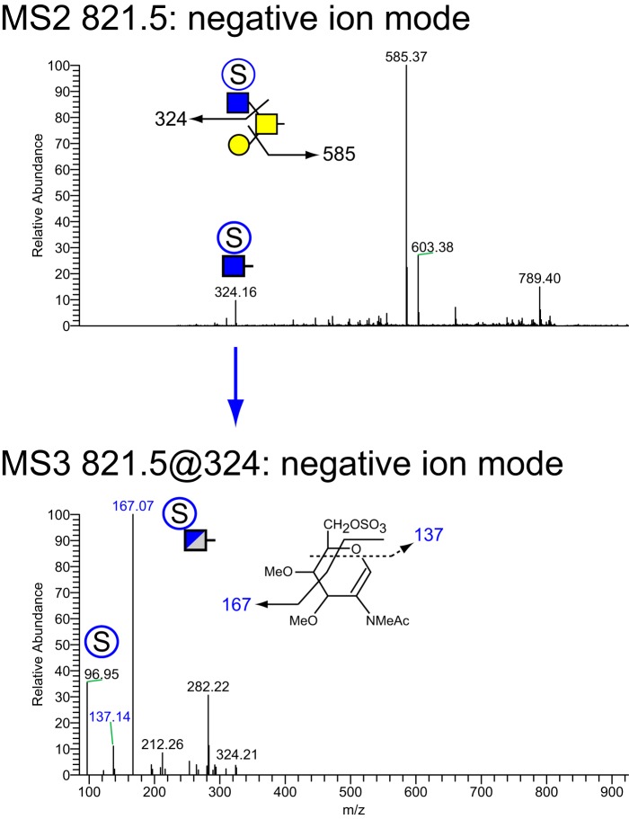
Figure 8.
NSI-MSn analysis of sulfated O-glycan structure 2. The glycan composition detected at m/z = 821.5 in negative mode (Table 1 and Fig. 7) was fragmented to determine the site and position of sulfation. Informative cross-ring fragments placed the sulfate at the 6-position of the branching HexNAc residue of this core 2 glycan.

