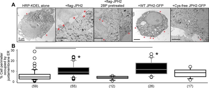
Figure 5.
Palmitoylation of JPH2 stabilizes ER–PM junctions assessed by quantifying juxtamembrane ER elements using transmission EM. COS-7 cells were transfected with horseradish peroxidase (HRP)-conjugated-KDEL, alone or with specified JPH2 variants. In the case of FLAG–JPH2, cells were incubated under the control conditions or in the presence of 2BP (100 μm) overnight before experiment. The ER lumen was marked by amplification of HRP–KDEL followed by peroxidase reaction with DAB in 0.01% H2O2 (16). A, representative TEM images. Red asterisks mark juxtamembrane ER. All scale bars refer to 5 μm. B, summary of percent cell perimeter occupied by juxtamembrane ER. Data were pooled from three independent experiments; numbers of cells analyzed are listed in parentheses. One-way ANOVA of all five groups, p < 0.001, followed by Dunn's tests versus HRP–KDEL alone; *, p < 0.05. Both FLAG–JPH2 and JPH2–GFP increased juxtamembrane ER elements. Inhibiting FLAG–JPH2 palmitoylation by 2BP or replacing all four Cys side chains with Ala in Cys-free JPH2–GFP nullified this effect.

