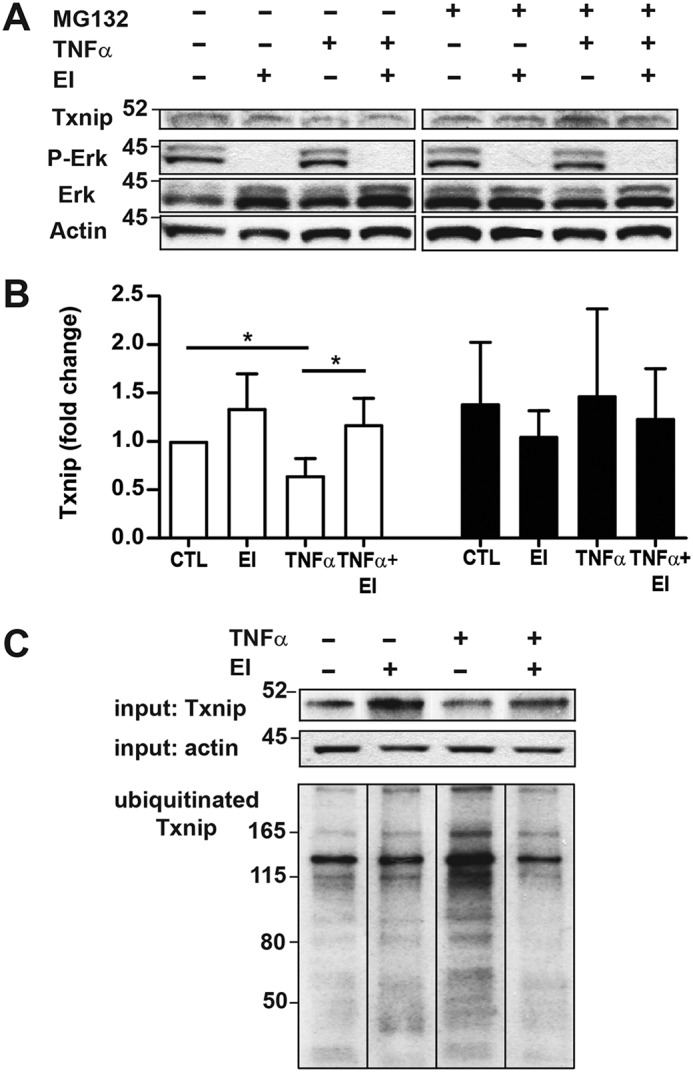
Figure 1.

Cytokine-induced degradation of Txnip is ERK- and proteasome-dependent. A549 cells were treated with the ERK1/2 MAP kinase inhibitor (EI) (U0126, 10 μm, 60 min) and the proteasome inhibitor MG132 (10 μm) and stimulated with TNFα (10 ng/ml, 60 min) followed by cell lysate preparation. A, immunoblots of Txnip, phospho-ERK (P-ERK), ERK, and β-actin. B, densitometric analysis of Txnip blots shown in A. Open bars, −MG132; filled bars, +MG132 (n = 3 per condition, one-sample t test). *, p < 0.05. C, A549 cells were treated with the proteasome inhibitor MG132 (10 μm) prior to ERK inhibition and TNFα stimulation. Ubiquitinated proteins were then isolated from protein lysates by IP, and Txnip immunoblots were prepared from IP eluates. CTL, control.
