Figure 4.

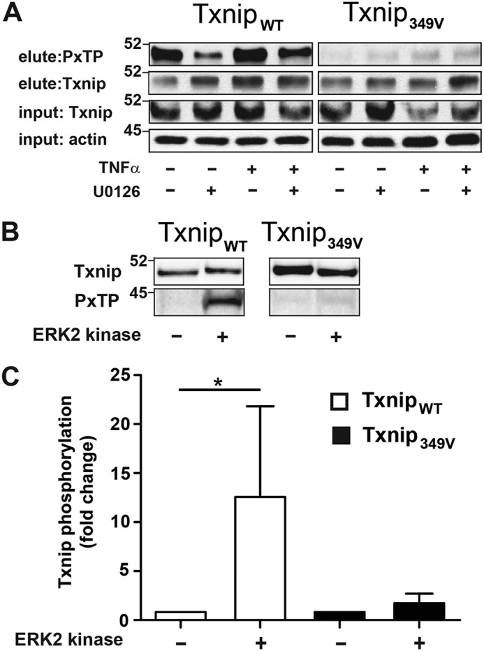
Cytokine-induced phosphorylation of Txnip threonine 349 (Thr349) by ERK. A, A549 cells were infected with lentivirus that express either C-terminally FLAG-tagged WT (TxnipWT) or T349V (Txnip349V) Txnip. The cells were treated with ERK1/2 MAP kinase inhibitor (U0126, 10 μm) and stimulated with TNFα (10 ng/ml, 60 min), and protein lysates were prepared. Txnip was immunoprecipitated with anti–FLAG-agarose, and immunoblots of eluate were probed with a phospho-MAPK (PXTP) and Txnip antibody. B, protein lysates were prepared from A549 cells expressing either TxnipWT or Txnip349V. Txnip was immunoprecipitated from cell lysate with anti-FLAG agarose and treated with ERK2 kinase (5 ng/μl), 60 min, 30 °C. PXTP and Txnip immunoblots were prepared from IP eluate. C, densitometric analysis of PXTP blots shown in B (n = 4 per condition, one-sample t test). *, p < 0.01.
