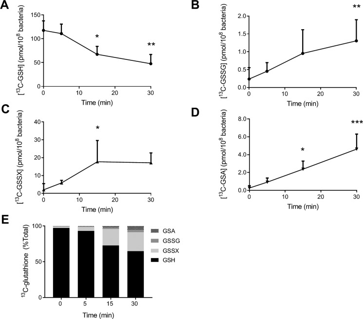
Figure 3.
Oxidation of GSH in P. aeruginosa during phagocytosis by human neutrophils. PAO1 (1 × 10⁸/ml) grown on CELTONE Complete 13C-medium was incubated with neutrophils (1 × 10⁷/ml) at 37 °C with end–over–end rotation. At the indicated time points, phagocytosis was stopped by placing the mixtures on melting ice; NEM (20 mm) was added, and neutrophils and bacteria were lysed. [13C]GSH (A), [13C]GSSG (B), [13C]GSSX (C), and [13C]GSA (D) were measured by stable isotope dilution LC-MS/MS. A significant difference when compared with time 0 was identified by ANOVA with Dunnett's multiple comparison test and is indicated by * (p < 0.05), ** (p < 0.01), or *** (p < 0.001). Data were obtained for at least three different donors and presented as mean ± S.D. E, individual glutathione species are presented as a percentage of total glutathione as described in Fig. 2. Data are presented as means from at least three independent experiments.

