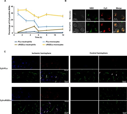
Fig. 2. The uptake of cRGDLs by M/Ns in vivo of I/R rats.

(A) The percent of M/Ns loading with Cy5-cRGDLs in blood circulation of I/R rats assayed by flow cytometer (n = 3). (B) Confocal microscope images of cellular location of Cy5-NBD-cRGDLs in isolated M/Ns from I/R rats. Red: Cy5, hydrophilic core marker and green: NBD-PE, lipid membrane marker. (C) Confocal microscope images of comigration of Cy5-PLs/Cy5-cRGDLs with M/Ns across cerebral vessels in the ischemic hemisphere 12 hours after reperfusion. Blue: M/Ns stained with cell tracer CFSE; red: Cy5-PLs or Cy5-cRGDLs; and green: cerebral vessels labeled with vWF rabbit anti-rat antibody and secondary antibody of Alexa Fluor 594 goat anti-rabbit.
