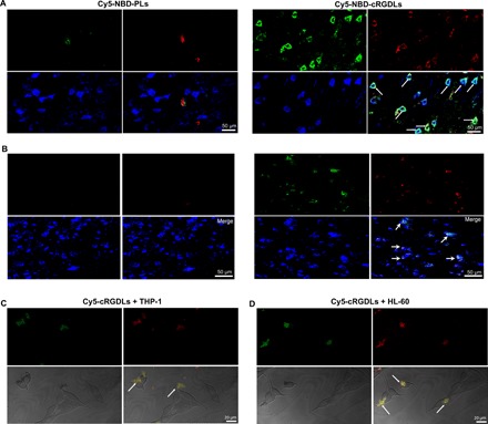
Fig. 4. The liposome transport from M/Ns to neuron in vitro and in vivo.

(A) Confocal microscope images of colocation of Cy5-NBD-PLs/Cy5-NBD-cRGDLs with neurons or microglia (B) in brains of I/R rats 12 hours after reperfusion. Green: NBD-PE marker; red: Cy5 marker; blue: neurons stained with Rabbit anti-rat MAP2 or mouse anti-rat Iba-1 antibody and secondary antibody of Alexa Fluor 405 goat anti-rabbit. (C) Confocal microscope images of the transfer of Cy5-cRGDLs from THP-1 or (D) HL-60 to PC12. Green: THP-1 or HL-60 stained with DiI; red: Cy5-cRGDLs; PC12 was not stained.
