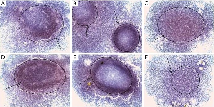
Figure 8.
Immunohistochemical staining of CSC markers by eMCTS cells transferred from the suspension culture to the adhesive culture of MCF-7 cells: (A) Bmi-1, (B) CD44, (C) CD133, (D) EpCAM, (E) vim, (F) CD24; hematoxylin, ×100 magnification, immunopositive cells [black arrows (PolyVueHRP/DAB Diagnostic BioSystems, USA)]. CSC, cancer stem cell2.

