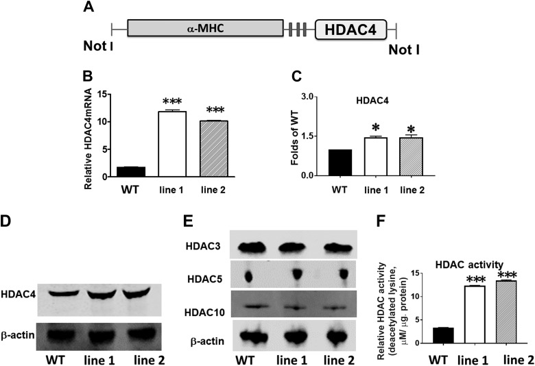
Fig. 1.
Generation and characterization of cardiac myocyte-specific histone deacetylase 4 (HDAC4)-transgenic (Tg) mice. A: schematic diagram of transgenic construct used to generate α-myosin heavy chain (α-MHC)-HDAC4 mice. B: quantitative PCR analysis of HDAC4 mRNA from the heart in wild-type (WT) and HDAC4-Tg mice. Results represent means ± SE (n = 5/group); ***P < 0.001. C: densitometric analysis shows the increased HDAC4 protein in HDAC4-Tg mice; *P < 0.05 vs. WT (n = 3/group). D and E: immunoblot detection of HDAC4 and other HDACs in WT and HDAC4-Tg mice. F: HDAC activity increased in HDAC4-Tg and wild type control myocardium; ***P < 0.001 vs. WT (n = 4/group). NotI (restriction enzymes).

