Fig. 2.
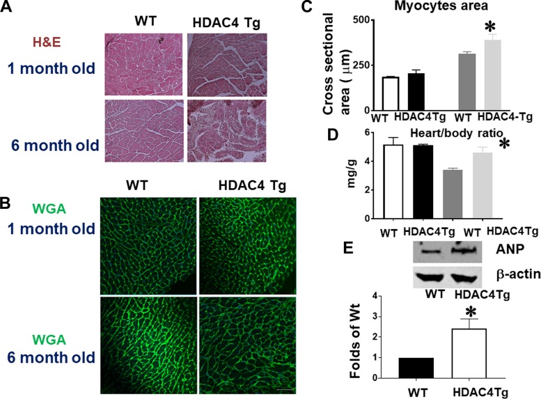
Histone deacetylase 4 (HDAC4) activation induced cardiac hypertrophy in the adult heart. A and B: HDAC4 overexpression increased myocyte sizes, as indicated by wheat germ agglutinin (WGA) and hematoxylin and eosin (H & E) staining (images taken at ×20 magnification). C: myocyte size from wild-type (WT) and HDAC4-transgenic (Tg) mice. D: heart/body ratios in WT and HDAC4-Tg mice (n = 3–5/group). E: atrial natriuretic peptide (ANP) protein levels in the myocardium of WT and HDAC4-Tg mice (n = 3/group). F: Masson’s trichrome staining indicates interstitial fibrosis in the myocardium (arrows indicate intensive fibrosis). G and H: Picrosirius red staining in WT and HDAC4-Tg mice and statistical analysis (images taken at ×20 magnification). I: immunostaining detection of CD31-positive microvessels from WT and HDAC4-Tg mice. Results represent means ± SE (n = 4–5/group). Scale bar, 50 μm. *P < 0.05 vs. WT; ***P < 0.001 vs WT.

