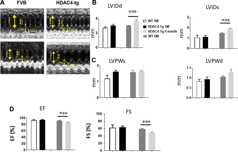
Fig. 3.
Echocardiographic measurements of ventricular function. Echocardiographic measurements of ventricular functional parameter at 1 and 6 mo age, A: representative images are shown as the M-mode short-axis ultrasound among the groups. B: left ventricle internal diameter at end diastole (LVIDd) and end systole (LVIDs). C: left ventricle posterior wall thickness at end diastole (LVPWd) and end systole (LVPWs). D: ejection fraction (EF) and fraction shortening (FS). Values represent means ± SE (n = 4–5/group); ***P < 0.01.

