Fig. 5.
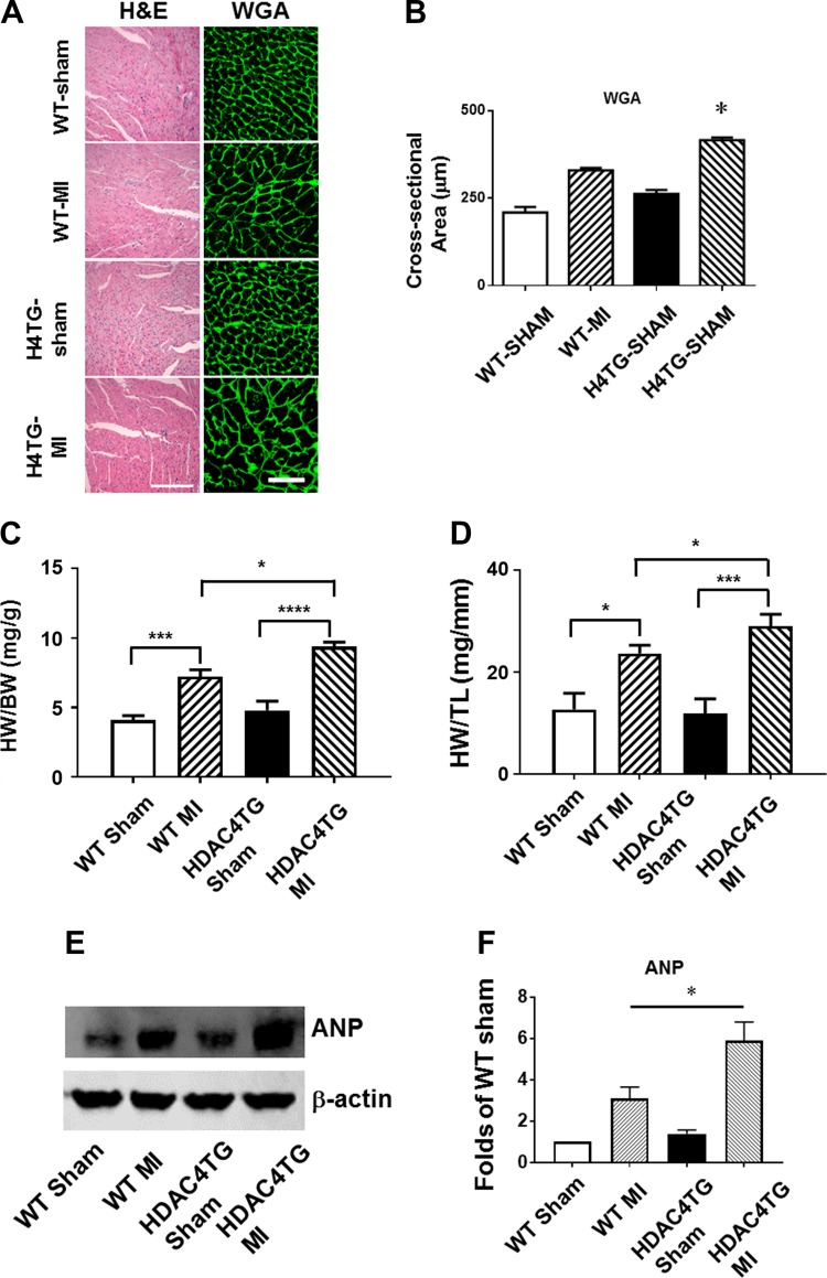
Histone deacetylase 4 (HDAC4) overexpression increased cardiac hypertrophy, interstitial fibrosis, and apoptosis. A and B: wheat germ agglutinin (WGA) and hematoxylin and eosin (H & E) staining showing the cross-sectional area of cardiomyocytes. C and D: heart weight/body weight (HW/BW) and heart weight/tibia length (HW/TL) ratio. Values represent means ± SE (n = 5/group). E and F: Western blot showing the increased atrial natriuretic peptide (ANP) protein in HDAC4-transgenic (Tg) mice in post-myocardial infarcted (MI) heart (n = 3/group). G and H: Picrosirius red staining showing the interstitial fibrosis of myocardium (n = 3/group). Scale bar, 200 μm. I: statistical analysis of apoptotic positive signals among the groups; J: representative images of TUNEL staining from the post-MI hearts of 2-mo-old mice (n = 5/group). Scale, 50 μm in A and 200 μm in J. *P < 0.05, ***P < 0.01, and ****P < 0.001 vs. MI.

