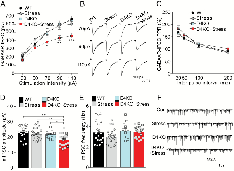
Fig. 4.
Stressed D4KO mice have the significantly diminished GABAergic synaptic inhibition in prefrontal cortex. (A, B, C) Input-output curves of GABAAR-IPSC (A), representative traces (B), and plot of paired-pulse ratio of GABAAR-IPSC (C) in prefrontal cortex (PFC) pyramidal neurons from wild-type (WT; n = 23 cells), Stress (n = 15 cells), D4KO (D4KO, n = 11 cells), and D4KO+Stress (n = 17 cells) mice. (D, E, F) Miniature IPSC amplitude (D), frequency (E) and representative traces (F) in PFC pyramidal neurons from WT (n = 21 cells), Stress (n = 24 cells), D4KO (n = 12 cells), and D4KO+Stress (n = 22 cells) mice. *P < .05, **P < .01, ***P < .001; 2-way ANOVA.

