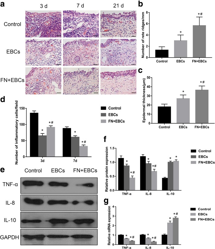
Fig. 4.
Histological features and expression of inflammatory factors of the rats’ dorsal wounds in each group. a Wound tissue sections stained with H&E on post-injury days 3, 7, and 21 showing histological features in rats’ dorsal wound treated with PBS (control), EBCs, and FN + EBCs. b, c Analyses of rate ridges numbers and epidermal thickness of wound tissue sections treated with PBS (control), EBCs, and FN + EBCs on post-injury day 21 showing histological features in rats’ dorsal wound. FN + EBCs treated wound displayed significantly more rate ridges and thicker epidermis than the others. d The number of inflammatory cells on day 3 and day 7 was quantified at per × 40 magnification for five areas randomly, FN + EBCs treated wound displayed significantly lower inflammatory response and fewer inflammatory cells on day 7. e, f Representative western blot and results of densitometric analysis of blots showing relative protein levels of TNF-α, IL-8, and IL-10 for each group on post-injury day 7. g Representative qRT-PCR analysis showing relative mRNA levels of TNF-α, IL-8, and IL-10 for each group on post-injury day 7. The values were analyzed by Graph Prism 7.0. Error bars represent SEM. Student’s t test, *P < 0.05 compared with control value (n = 6), #P < 0.05 compared with EBCs value (n = 6). Scale bar, 100 μm

