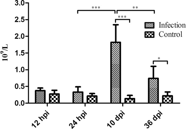
Fig. 1.

The number of eosinophils in the infected and control groups at the four indicated infection stages in the blood of Beagle dogs infected with 300 T. canis embryonated eggs were analyzed by t-test using GraphPad Prism v.5. [24 h infection group vs 10 d infection group: t(11) = 6.64, P < 0.001; 10 d infection group vs 10 d control group: t(11) = 7.67, P < 0.001; 10 d infection group vs 36 d infection group: t(11) = 4.22, P < 0.01; 36 d infection group vs 36 d control group: t(10) = 3.37, P < 0.05]. *P < 0.05, **P < 0.01, ***P < 0.001
