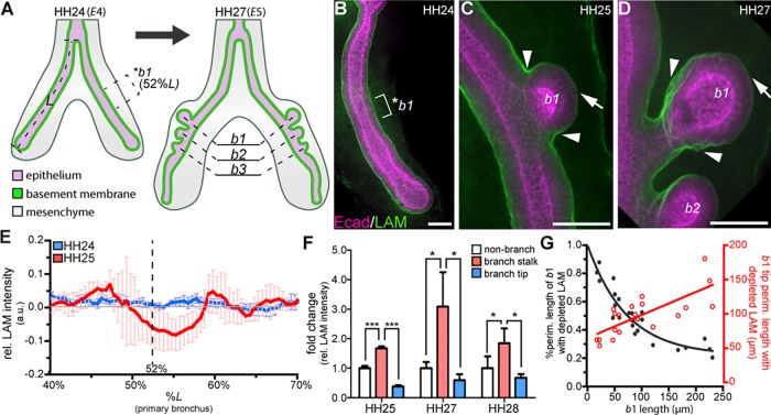
Fig. 3.
Basement membrane thinning occurs after branch initiation and during branch extension. (A) Schematic of embryonic chicken lung showing the stereotyped position at which b1 initiates (52%L, where L is length from the tracheal fork to the distal tip of the primary bronchus). (B-D) Staining for the basement membrane protein laminin (LAM, green) and for E-cadherin (Ecad, magenta) in the airway epithelium (B) prior to b1 initiation, (C) during b1 initiation and (D) during b1 extension. Laminin intensity increases at branch stalk (arrowheads, C,D) and decreases at branch tip (arrows, C,D). *b1 indicates the region (brackets) where b1 will form. (E) Plot of laminin intensity at b1 before (blue, HH24) and during (red, HH25) branch initiation; n=3-5 independent experiments. (F) Mean laminin staining intensity in the branch stalk (arrowheads in C,D) and branch tip (arrows in C,D) normalized to non-branching regions; n=3 independent experiments. (G) Plot of the percentage of cross-sectional b1 surface (black) and the size of the b1 surface (red) that shows depletion of laminin as a function of b1 length; n=20 lung explants. Data are mean±s.d. *P<0.05, ***P<0.001 using one-way ANOVA with Tukey's post-hoc test. Scale bars: 40 µm.

