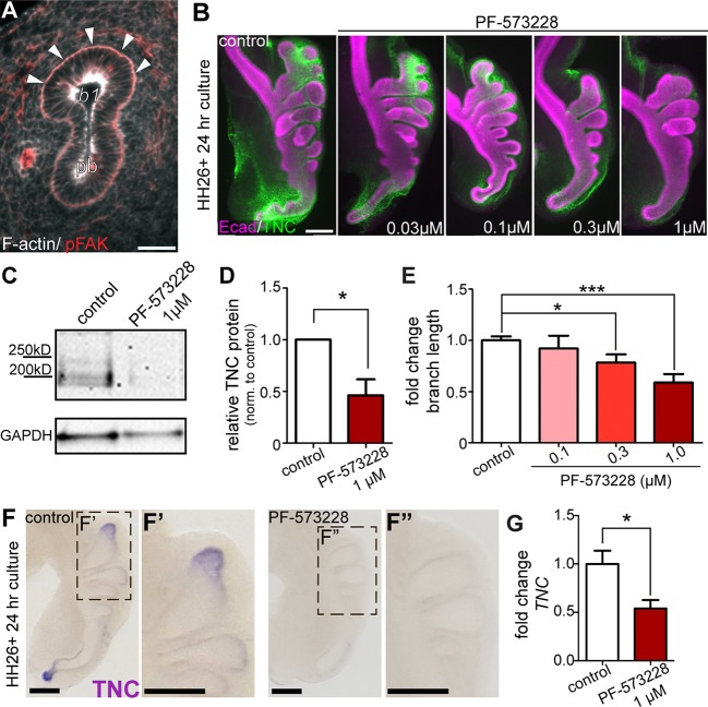
Fig. 6.
FAK is required for TNC expression and branch extension. (A) Histological section shows pFAK (red, arrowheads) localized to the basal surface of the airway epithelium. (B) Explants isolated at HH26 were cultured for 24 h in the presence of increasing concentrations of the FAK inhibitor PF-573228. TNC distribution around airway epithelial branches was observed with immunostaining (TNC, green; Ecad, magenta). Explants cultured in the presence of PF-573228 were compared with untreated controls. (C) Immunoblotting for TNC was performed on protein extracts isolated from HH26 lung explants cultured for 24 h and (D) fold change of TNC band intensity was quantified in PF-573228-treated explants relative to control; n=3 independent experiments. (E) Fold-change in branch extension of explants treated with increasing concentrations of PF-573228 for 24 h; n=4 or 5 independent experiments. (F-F″) In situ hybridization for TNC transcript in control and PF-573228-treated lung explants. (G) qRT-PCR analysis of explants cultured in the presence of PF-573228 for 24 h; n=3 independent experiments. Data are mean±s.d. *P<0.05, ***P<0.001 using (D,G) Student's t-test or (E) one-way ANOVA with Tukey's post-hoc test. Scale bars: 40 µm in A; 200 µm in B,F.

