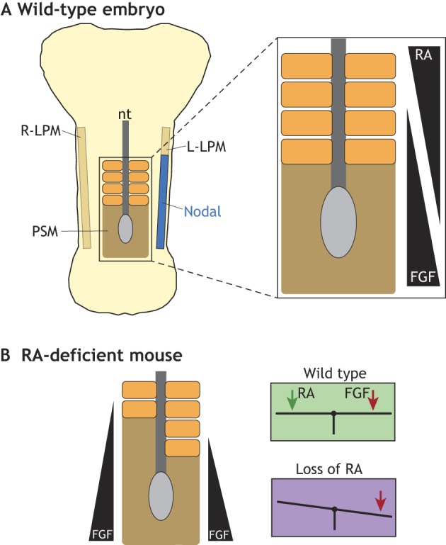
Fig. 5.

The maintenance of symmetry during segmentation. (A) Schematic of a somite-stage mouse embryo showing the lateral plate mesoderm (LPM) with left-sided L-R pathway activity (blue), the central neural tube (nt) and the paraxial mesoderm (orange), the unsegmented region of which is called the pre-somitic mesoderm (PSM, brown). Opposing gradients of RA and FGF activity determine the position of the determination front, which is the position at which cells initiate the segmentation program. (B) In the absence of RA, mouse embryos exhibit asymmetric somites, with the right side showing a delay owing to right-biased FGF8 activity. This results from the loss of buffering by RA, which acts as a contralateral balancer to the somite desynchronizing activity of FGF8.
