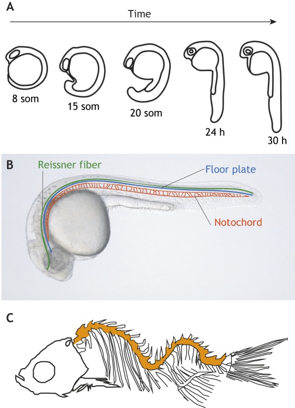
Fig. 6.

Zebrafish body symmetry depends on motile cilia-dependent mechanisms. (A) Schematic of the first 30 h of zebrafish development. Initially, during somite (som) stages, zebrafish embryos are curved around the yolk ball. As the body axis extends, the trunk moves dorsally to straighten this initial curve. (B) Image of a 30 h zebrafish embryo schematically showing the position of the Reissner fiber, the floor plate of the neural tube and the underlying notochord. (C) Schematic skeleton of a mutant zebrafish in which cilia motility is inactivated. Mutants exhibit three-dimensional spinal curves (orange) that resemble aspects of idiopathic scoliosis.
