
Fig. 5.
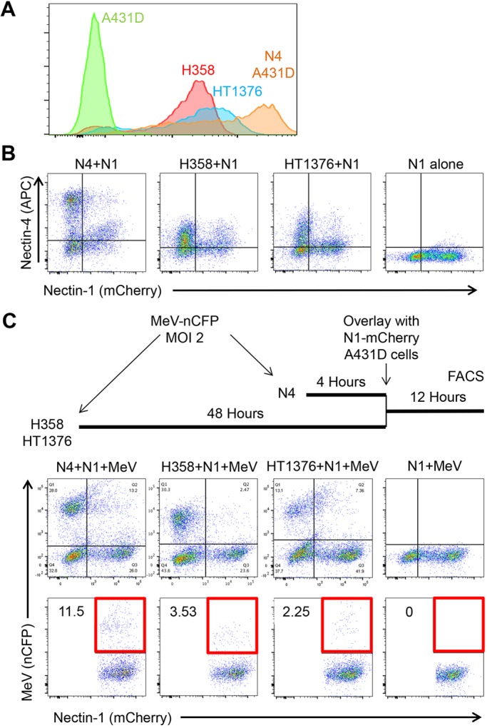
Epithelial cell lines expressing physiological N4 levels can spread infection. (A) FACS analysis of N4 expression levels in epithelial cell lines H358 and HT1376, as compared to N4–Dendra2 A431D cells. Primary antibody 337516 (R&D Systems) was used to detect N4, with a secondary antibody emitting fluorescence in the PE channel. (B) N1–mCherry-expressing cells internalize N4 from H358 and HT1376 cells. N1–mCherry A431D cells were overlaid on N4–Dendra2-expressing A431D cells (first panel), H358 cells (second panel) or HT1376 cells (third panel), or plated alone (fourth panel), incubated for 12 h, fixed, permeabilized, antibody stained and analyzed by FACS. Antibody 337516 was used to detect N4, with a secondary antibody emitting fluorescence in the APC channel. (C) N1–mCherry expressing cells can internalize MeV RNP from H358 and HT1376 cells. Top: schematic drawing of the experiment. Center row of panels: nCFP expression in mixed cultures infected by MeV–nCFP. Panel order is as in B. Bottom row of panels: quantitative analysis of the double-positive (N1-mCherry and nCFP) cells. The areas selected for quantification are indicated by a red square.