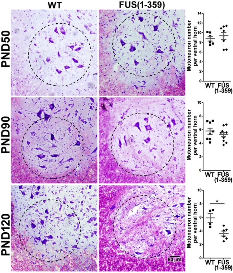
Fig. 1.

Nissl staining revealed significant motor neuron loss by P120 in FUS (1-359) mice. Nissl stain highlights nucleic acid, in particular ribosomal RNA, which is abundant in motor neurons and results in a dark purple stain of the cell body. Figure shows representative images from the ventral horn area of spinal cord tissue from WT and FUS (1-359) transgenic mice, accompanied by the quantification scatter-plot graph, obtained at P50 [top; WT n=6, FUS (1-359) n=8], P90 [middle; WT n=8, FUS (1-359) n=10] and P120 [bottom; WT n=4, FUS (1-359) n=6]. Dashed circle indicates ventral horn region used for quantification. Each datapoint represents mean motor neuron counts from 20 non-consecutive tissue slices along the lumbar spinal cord, dark grey lines represent mean±s.e.m. *P=0.005 [t-test: WT versus FUS (1-359) at P120].
