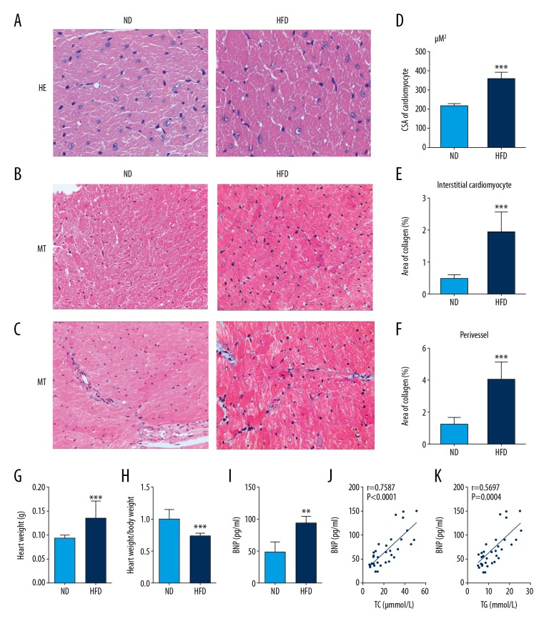
Figure 2.
A high-fat diet (HFD) increased cardiac hypertrophy and myocardial fibrosis in C57BL/6J mice. (A) Representative photomicrographs of the light microscopy show the cross-sectional area (CSA) in cardiomyocytes. Hematoxylin and eosin (H&E). Magnification ×400. (B, C) Representative photomicrographs of the light microscopy show myocardial fibrosis in the left ventricles. Masson’s trichrome. Magnification × 200. (D–F) Quantitative analysis of cardiomyocyte CSA, and the percentage area of collagen in the cardiac interstitium and around myocardial vessels in the left ventricle. (G, H) HFD is associated with a significant increase in heart weight and levels of brain natriuretic peptide (BNP). (I, J, K) BNP is positively correlated with serum lipid levels in mice (analyzed by Spearman correlation). Results are presented as the mean ±SD. * P<0.05, ** P<0.01, *** P<0.001 vs. ND group.

