Fig. 1.
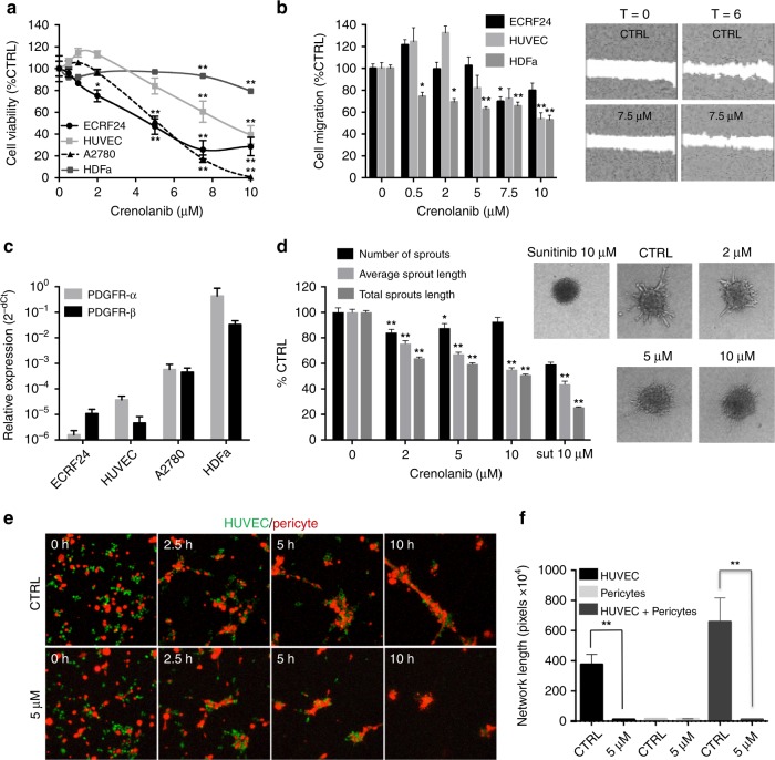
Activity of crenolanib on cell viability, migration and sprouting. a Cell viability dose response curves of crenolanib in endothelial cells (immortalised ECRF24 and primary human umbilical vein endothelial cells (HUVEC), ovarian cancer cells (A2780) and adult human dermal fibroblasts (HDFa). Cell viability was assessed after 72 h of exposure to crenolanib and represented as a percentage of untreated controls. Significance is indicated versus untreated cells. b Endothelial cell migration in response to crenolanib. Cell migration was assessed after 6 h drug treatment using a scratch assay. c PDGFR-α and -β expression in ECRF24, HUVEC, A2780 and HDFa determined by qPCR. d Activity of crenolanib on HUVEC sprouting. The number of sprouts and the average sprout length were quantified. e Representative images of HUVEC (green) and human pericyte (red) co-cultures. Co-cultures were established in 3D Matrigel matrices and allowed to randomly co-assemble over 10 h in the presence of DMSO or crenolanib (5 µM) at 0, 2.5, 5 and 10 h. f Quantification of the network length of capillary-like structures in HUVEC alone, pericyte alone and HUVEC/pericyte co-culture in the presence or absence of crenolanib (5 µM) at 10 h. All values shown are presented as percentage of the CTRL and represent the mean of at least two experiments performed in triplicate. Cells treated with 0.1% DMSO were used as a control (CTRL). Error bars indicate SEM. Significance (*P < 0.05, **P < 0.01) is indicated as compared to control

