Fig. 2.
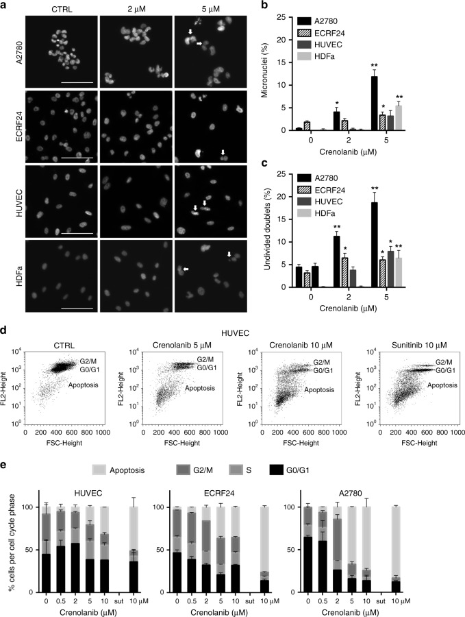
Crenolanib induces nuclear aberrations and apoptosis. a DAPI staining of A2780, ECRF24, HUVEC and HDFa after treatment with crenolanib for 72 h. In cells treated with crenolanib 5 µM white arrowheads indicate either micronuclei or undivided nuclear doublets. b, c Quantification of the percentage of micronuclei (b) and undivided nuclear doublets (c) based on images of DAPI staining. Values shown represent the percentage aberrations of the total amount of cell nuclei per image field. d Propidium-iodide staining of HUVEC treated with crenolanib for 72 h using flow cytometry. Cellular DNA content in permeabilised cells is proportional to fluorescence intensity (FL2-H; y-axis) and allows for the distinction of cells containing diploid DNA (G0/G1), tetraploid DNA (G2/M) and subdiploid DNA (apoptotic fraction). e Quantification of cellular DNA distribution over cell cycle phases as indicated in HUVEC, ECRF24 and A2780 cells after crenolanib treatment. All values shown are presented as percentage of the CTRL and represent the mean of at least two experiments. Error bars indicate SEM. Significance (*P < 0.05, **P < 0.01) is indicated as compared to control

