Fig. 1.
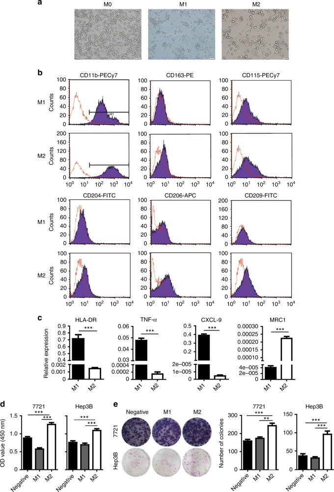
Polarisation of THP-1 to M1-like and M2-like macrophages. a Cell morphology of M0-like, M1-like, or M2-like macrophages. b Macrophage marker expressions of M1-like and M2-like macrophages were detected by flow cytometry assay using various specific antibody (black histograms) or isotype-matched antibody (red histogram). c Expressions of HLA-DR, TNF-α, CXCL-9 and MRC1 in M1 or M2 macrophages was determined by RT-qPCR. d Effects of M1-CM and M2-CM on the proliferation (at 48 h) of SMMC-7721 (left) and Hep3B cells (right), respectively. e Effects of M1-CM and M2-CM on the colony formation of SMMC-7721 and Hep3B cells respectively. N = 3, mean ± SEM, **P < 0.01, ***P < 0.001

