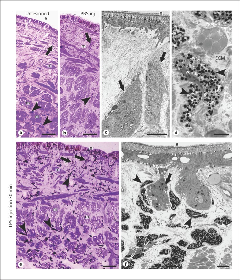
Fig. 2.
Morphological analysis of leech body wall at optical and TEM microscopes 30 min after PBS and LPS injection. Few resident macrophages (arrow) and vasofibrous tissue cells (arrowheads) are visible underneath the epithelium (e) or among the muscle fibres (m) in unlesioned (a) and in PBS-injected animals (b). Detailed TEM of macrophages (arrows in c) and of the vasofibrous tissue (d) surrounded by extracellular matrix (ECM) formed by vasocentral cell (vc), with cytoplasm containing a few granules, surrounded by vasofibrous cells (arrowheads), with a cytoplasm containing numerous small highly electron-dense granules. 30 min after LPS injection (e, f), numerous vasofibrous tissue cells are recognizable by their dark color (arrowheads in e) among muscle fibers and underneath the epithelium (e). f Detailed view of type I granulocytes (arrowheads) detached from vasocentral cells (vc) and next to resident macrophages (arrow) localized in the subepithelial region (e). Bars in a, b, e: 100 μm; bar in c, d: 2 μm; bar in f: 10 nm.

