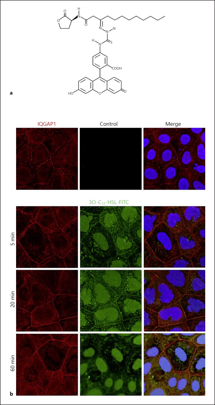
Fig. 4.
Imaging of IQGAP1 and 3O-C12-HSL in human intestinal epithelial cells. a Synthetic fluorescently tagged 3O-C12-HSL-FITC used for imaging. b Caco-2 human intestinal epithelial cells were treated with 1 µM of 3O-C12-HSL-FITC (green) or 0.018% DMSO as a diluent control for 5, 20, or 60 min. Samples were fixed, and then the IQGAP1 was immunolabelled (Atto647N; red) and the nucleus stained with DAPI (blue), as described previously [25]. The samples were visualized by confocal imaging. The image size is 67.6 × 67.6 µm and pixel size is 0.13 µm.

