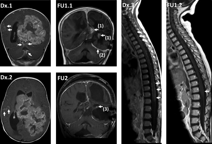
Figure 3.
Course of magnetic resonance imaging (MRI) in patient 4, reaching a complete remission without radiotherapy. At diagnosis, cerebral MRI (cMRI) coronal (cor) enhanced T1‐weighted image (T1 CE) (Dx.1) and axial T1 CE (Dx.2) at diagnosis showed large primary tumor in the left hemisphere, crossing the midline and reaching the lateral ventricles, with extensive intracranial leptomeningeal dissemination (arrows), and spinal MRI (spMRI) sagittal (sag) T1 CE (Dx.3) showed extensive spinal leptomeningeal dissemination (arrows). One year after diagnosis, after previous local progression on primary treatment, reoperation, and individual salvage chemotherapy, cMRI cor T1 CE (FU1.1) showed small residual tumor [arrows (1)] and no residual leptomeningeal seeding (partial response). Typical postinterventional subdural enhancement [arrow (2)]. Also 1 year after diagnosis, spMRI sag T1 CE (FU1.2) showed good response with minimal leptomeningeal dissemination [arrow]. Eight years after diagnosis, cMRI cor T1 CE (FU2) showed residual contrast enhancing tissue [arrow (3)]. After initial shrinking, the aspect had been without any change over the last years with regard to size or contrast enhancement and was regarded as gliotic tissue (complete remission). Based on the stability of imaging aspect, histological verification of complete remission was not pursued.

