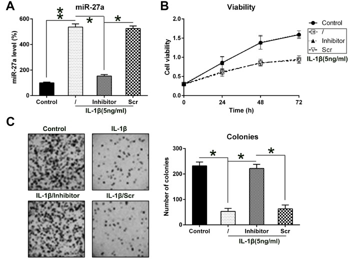
Figure 3.
Contribution of miR-27a expression to the growth of IL-1β-triggered chondrocytes. (A) Expression levels of miR-27a in IL-1β-treated chondrocytes that underwent transfection with the miR-27a inhibitor and miR-Scr were evaluated by Q-PCR. (B) The proliferation rate of IL-1β-treated chondrocytes was measured at 24, 48, and 72 h following their transfection using the MTT test. (C) Soft agar colony generation assay of IL-1β-treated chondrocytes transfected with the miR-27a inhibitor and miR-Scr. The lower panel indicates the number of colonies in each group. The results are described as the mean ± SD. *P < 0.05, **P < 0.01 vs. indicated group.

