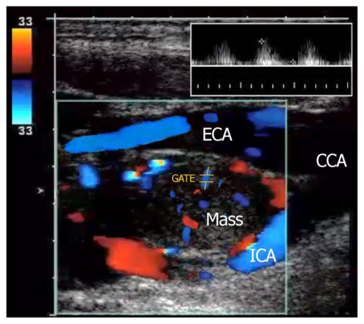
Figure 2.
42-year-old female with a left carotid body tumor.
FINDINGS: Doppler ultrasound shows a well-defined, hypoechoic mass locates at the carotid bifurcation between the internal (ICA) and external carotid artery (ECA). Duplex imaging in the same patient shows the highly vascular nature of the paraganglioma located in the crux of the bifurcation. Waveform analysis (inset with sampling location is GATE) shows the internal arterial flow of this lesion which demonstrates low-resistance artery characteristics.
TECHNIQUE: Aloka F37, 7.5 MHz linear probe, Doppler ultrasonography.

