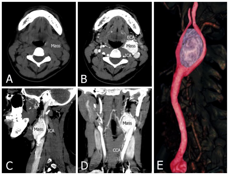
Figure 3.
42-year-old female with a left carotid body tumor.
FINDINGS: (A) Non-contrast CT shows a mass with soft tissue density. (B, C, and D) Contrast-enhanced CT with early arterial phase contrast shows a heterogeneously enhancing mass at the left carotid bifurcation. This lesion has bright and rapid enhancement. It demonstrates a carotid body tumor splaying the bifurcation of the internal (ICA) and external (ECA) carotid arteries. (E) CT angiography 3D reformat image of left carotid body tumor.
TECHNIQUE: Philips 64 CT scanner with non-contrast and contrast, 500 mA, 120 kVp, Slice thickness = 1.25 mm, a total of 60 mls of Omnipaque 350 was administered intravenously.
a) Axial soft tissue window, 1.25 mm slice thickness, non-contrast CT
b) Axial soft tissue window, 1.25 mm slice thickness, contrast CT
b) Sagittal soft tissue window, 1.25 mm slice thickness, contrast CT
c) Coronal soft tissue widow, 1.25 mm slice thickness, contrast CT
e) CT angiography 3D reformatted image, slice thickness = 15 mm.

